Intrasurgical OCT EnFocus
See subsurface details to make insightful surgical decisions for your patient, with real-time, intrasurgical Optical Coherence Tomography (OCT).
A top-down microscope view, combined with your experience, helps you assess intraoperative changes to subsurface tissues. But what if you could supplement this with a real-time, cross-sectional view?
EnFocus intrasurgical OCT provides real-time imaging of ocular tissue microstructures with the high resolution and deepest scan depth of any available intrasurgical OCT. This additional information can aid your intrasurgical decision-making in cornea, glaucoma, and retina procedures. For example in membrane peel cases, OCT findings were not in line with the surgeons first impression in 35% of cases according to a recent study.*
Combine EnFocus with a Proveo 8 or M844 microscope for comprehensive visualization. Choose Proveo 8 for the optimal OR workflow solution.
OCT: From clinical assessment to intraoperative decisions
Bridge the gap between pre-operative assessment and real-time evaluation of changes to tissue microstructures during surgery with EnFocus OCT.
In a clinical setting, OCT has become the standard of care. OCT technology is now moving more and more into the operating room where it is impacting surgical decision-making. The DISCOVER* study using EnFocus showed that “in membrane peeling procedures, intraoperative OCT findings were discordant from the surgeon’s initial impression in 35% cases”
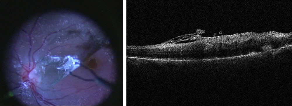
Benefits for your retina surgery
Use OCT to assess the level of tension in a membrane peel in real time, in order to avoid potential tears and protect the integrity of underlying tissue. A high-resolution view of <=4 µm also aids examination of retinal morphology for residual membranes and complications such as macular holes, or sub-retinal edema.
Additional benefits for posterior surgery include:
- Dynamic scan control via footswitch for swift adjustment of the scan angle to align with the membrane tissue
- Easy integration of fundus viewing systems such as the BIOM5 from OCULUS with synchronized focus
Benefits for your cornea surgery
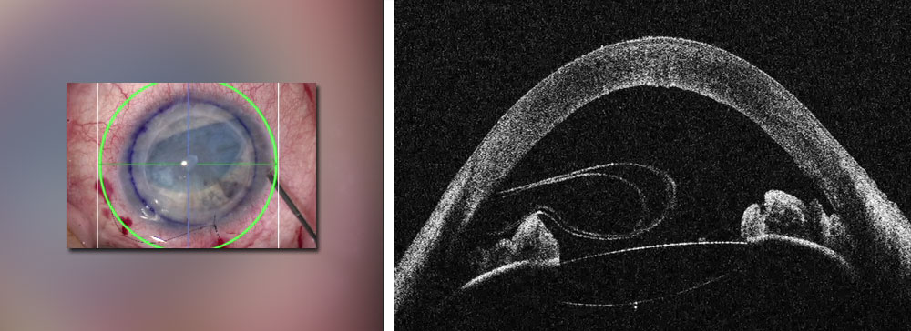
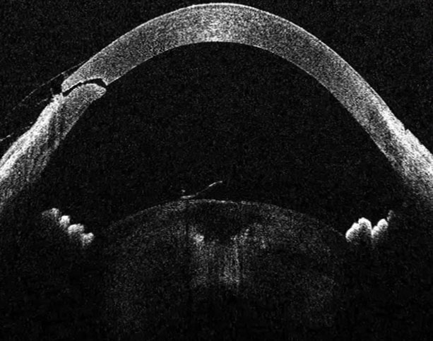
Easily view the entire anterior segment with the EnFocus Ultra-Deep system which has an increased depth of up to 11 mm.
In advanced lamellar corneal surgeries such as DMEK (Descemet’s Membrane Endothelial Keratoplasty) and DSEAK (Descemet’s Stripping Endothelial Automated Keratoplasty), this aids the surgeon in confirming the correct orientation of donor tissue which may help avoid corrective follow-up surgery.
Benefits for your glaucoma surgery
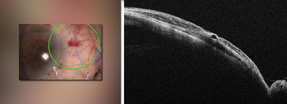
OCT scans of up to 20 mm wide and 11 mm deep support your view to aid accurate placement of an XEN gel stent.
OCT can also support shunt vessel placement and assessment of how much the tube should be tied off to control intraocular pressure. This can prevent further progression of the glaucoma.
EnFocus Ultra-HD OCT: Rich subsurface detail
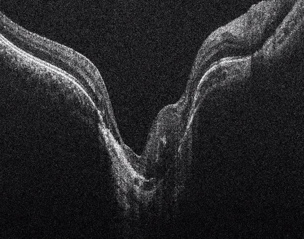
The best images matter. EnFocus Ultra-HD OCT technology delivers high definition real-time images of both the posterior and anterior segment.
Enfocus Intrasurgical OCT features:
- Resolution: ≤ 4 μm
- Depth: 2.5 mm image depth in tissue
- High OCT scan density (up to 1 million A-scans per volume)
EnFocus Ultra-Deep OCT: Deeper & wider
The EnFocus Ultra-Deep OCT option delivers very deep and wide imaging for full anterior segment visualization. Anterior structures continue to be resolved with crisp detail of ≤ 9 μm making it an ideal option for glaucoma surgery.
Enfocus Intrasurgical OCT features:
- Resolution: ≤ 9 μm
- Depth: 11 mm in tissue
- > 20 mm scan length
Efficiency you can feel, precision you can trust
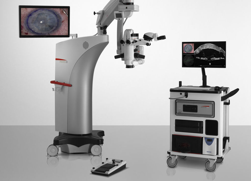
The EnFocus system combines perfectly with the Proveo 8 microscope for a comprehensive visualization system that is intuitive and straightforward to use.
- Easily control EnFocus OCT and microscope functions via integrated wireless footswitch
- Inject OCT images into the eyepieces and simultaneously display on the large HD monitor for smooth workflow
- Choose the Proveo 8 floor stand or ceiling-mount configurations for flexibility in your OR
- Smart workflow features and optical technologies mean that the Proveo 8 provides an optimal view at every stage of surgery, for interruption-free working
Simply capture and analyze
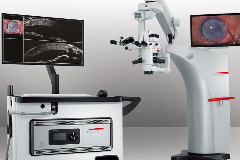
Reliably and efficiently obtain and view the information you need. Intuitive software supports your workflow with exactly the information and tools you need.
- Wide OCT viewing windows
- On-screen procedural pre-set modes
- Fully customizable scan management and dynamic scan control to adapt scan angle to membrane tissue
On-screen caliper measurements - Choose the full on-screen menu or switch to a simplified view for ease of use during your procedure
Display and record in HD
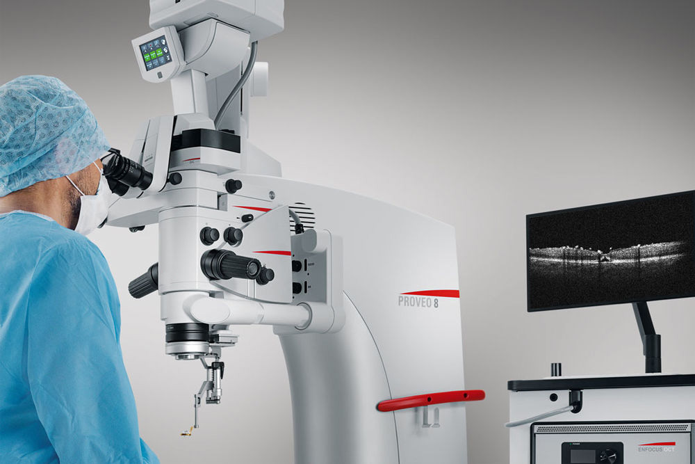
EnFocus captures the highest resolution images, then displays and records these in rich detail.
- Combine with the Proveo 8 microscope for real-time image injection into the eyepieces
- Record your surgery in full-HD for later review with just a tap of the footswitch or a click of the mouse
- Share the real-time view with your team via the large HD display
